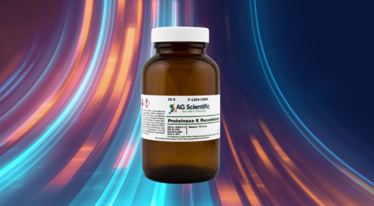
Proteinase K (also known as Endopeptidase K or Protease K) is a broad-spectrum serine protease that is able to digest native keratin (hair). It was discovered in extracts of the fungus Engyodontium album in 1974. The enzyme primarily cleaves the peptide bond adjacent to the carboxyl group of aliphatic and aromatic amino acids with blocked alpha amino groups.
Why is this enzyme used in DNA extraction?
Proteinase K is used during DNA extraction to digest many contaminating proteins present. It also degrades nucleases that may be present in DNA extraction and protects the nucleic acids from nuclease attack.
What are the applications of Proteinase K?
Applications for Next Generation Sequencing (NGS) and Microarray Technologies:
- Nucleic acid purification by inactivating nucleases when extracting DNA and RNA from yeast, bacteria, and mammalian cell & plant cell lysates
- Improves cloning efficiency of PCR products
- Sample preparation for quantification of DNA adduct levels by accelerator mass spectrometry
- Inactivation of enzyme cocktails in ribonuclease protection assays
- Added to extraction procedures to optimize RNA yields from primary breast tumors for microarray studies
Applications for Molecular Biology:
- Detection of Bovine Spongiform Encephalopathy proteins which are uniquely resistant to proteolytic degradation.
- Tissue digestion (denatures proteins) as an alternative sample preparation approach for quantitative analysis using liquid chromatography—tandem mass spectrometry.
- Specifically modify cell surface proteins to analyze membrane structures for protein localization
- Generates protein fragments used in characterization of functional studies
What are the guidelines for using PK?
- Isolation of high molecular weight DNA: Chromosomal DNA that has been embedded in agarose plugs can be treated with proteinase K to inactivate rare-cutting restriction enzymes used to digest the DNA. The enzyme is used for this method at a concentration of 1 mg/ml in a buffer containing 0.5M EDTA and 1% N-lauroylsarcosine (v/v). Incubate 24-48 hours at 37°C.
- Isolation of plasmid and genomic DNA: Genomic or plasmid DNA can be isolated from liquid nitrogen frozen cells or cultured cells using proteinase K. Incubate 50-100 mg of tissue or 1x10⁸ cells in 1 ml of buffer containing 0.5% SDS (w/v) with proteinase K at a concentration of 1 mg/ml, for 12-18 hours at 50°C.
- Isolation of RNA: For cytoplasmic RNA isolation, centrifuge the cell lysate, remove the supernate and add 200 ug/ml proteinase K and SDS to 2% (w/v). Incubate for 30 minutes at 37°C. Total RNA can be isolated by passing the lysate through a needle fitted to a syringe prior treatment with the enzyme.
- Inactivation of RNases, DNases and enzymes in reactions: Proteinase K is active in a wide variety of buffers. The enzyme should be used at a ratio of approximately 1:50 (w/w, proteinase K: enzyme). Incubation is at 37°C for 30 minutes.
Why is digestion performed at 50°C?
Increasing the temperature to 50°C will unfold some proteins making it easier for Proteinase K to degrade them. The enzyme is stable and activity is greatly increased with the addition of denaturing agents such as SDS and urea.
What is the quickest most effective way to inactivate proteinase K?
The most effective way to inactivate the enzyme, as with most proteins is to increase the temperature or change the pH significantly. Proteinase K is inactivated by heat (e.g. incubating at 55°C).
Activity in Buffers:
Buffer (pH 8.0, 50°C, 1.25 µg/ml protease K, 15 min incubation) | Proteinase K activity (%) |
30 mM Tris·Cl | 100% |
30 mM Tris·Cl; 30 mM EDTA; 5% Tween 20; 0.5% Triton X-100; 800
mM GuHCl |
313% |
36 mM Tris·Cl; 36 mM EDTA; 5% Tween 20; 0.36% Triton X-100; 735
mM GuHCl |
301% |
10 mM Tris·Cl; 25 mM EDTA; 100 mM NaCl; 0.5% SDS | 128% |
10 mM Tris·Cl; 100 mM EDTA; 20 mM NaCl; 1% Sarkosyl | 74% |
10 mM Tris·Cl; 50 mM KCl; 1.5 mM MgCl2; 0.45% Tween 20; 0.5%
Triton X-100 |
106% |
10 mM Tris·Cl; 100 mM EDTA; 0.5% SDS | 120% |
30 mM Tris·Cl; 10 mM EDTA; 1% SDS | 203% |
How do you determine if the enzyme is working?
To determine if the enzyme is working, you can do the following 2 steps:
-
- Find how many micromoles of the p-nitroanilide are produced per minute.
- Then divide by the total amount of protein in the solution, you can determine the specific activity of the enzyme = units (one unit equals 1 mole of p-nitroanilide produced/min), specific activity = units of enzyme activity/mg total protein.
Where does Proteinase K cleave?
Proteinase K cleaves peptide bonds next to the carboxyl group of N-substituted hydrophobic, aliphatic, and aromatic amino acids. It also cleaves peptide amides.
How long does this product last?
It has a shelf life of 12 months when stored in a dry place at 4–8°C, due to the fact that it is a very stable. Short term storage at ambient temperatures do not harm the enzyme’s activity and stability.
Download Proteinase K Brochure
Additional Reading
- 8 Benefits of Choosing AGS as Your Proteinase K Supplier
- Proteinase K - Bulk Packaging & Labeling Services
- 6 Reasons to Initiate Change Control with Manufacturers
We highly recommend storing PK at 4 degrees Celsius if you plan for long-term storage, but leaving PK out for a few days at room temperature should not ruin the reagent.
Thanks for commenting!
Best,
Trisha Timpug
Co-Director of Marketing and Sales
AG Scientific
Best wishes, Hayley
Inactivating proteinase K is perhaps one of the most common questions we see. And the answer is very simple. Heat is a widely used way of inactivating proteinase K. While the activity of proteinase K increases with temperature, and is optimized at about 65 ËšC, heating proteinase K to 95 ËšC for 10 minutes will inactivate it. Keep in mind, however, that heating proteinase K does not fully inactivate the enzyme. There will always be a small amount of activity remaining through this method.
Protease inhibitors such as PMSF and AEBSF (Pefabloc®) can also be used to permanently inactivate proteinase K.
Note- the actual inactivation temperature has been debated, ranging between 70 - 95 ËšC. However, crowd sourced feedback and extensive research led us to settle on 95 ËšC as the best temperature for inactivation.
Hope above information helps you.
Thank you,
Veera
Director of Web Content
Thank you for reading!
Idelle Delapena
Marketing Director
AG Scientific
https://www.researchgate.net/post/What_is_the_best_method_for_RNA_extraction_from_formalin-fixed_paraffin_wax_embedded_tissue_blocks
https://www.researchgate.net/topic/rna_isolation
Thank you!
Idelle Delapena
Marketing Director
You can follow any protocol mentioned in below links according to the sample you are using.
http://clinchem.aaccjnls.org/content/50/5/975.full
http://www3.appliedbiosystems.com/cms/groups/mcb_support/documents/generaldocuments/cms_041236.pdf
https://www.promega.com/~/media/files/resources/protocols/product%20information%20sheets/n/proteinase%20k%20protocol.pdf
http://www.ncbi.nlm.nih.gov/pubmed/21374178
Please let us know if you have other questions.
Thank you!
Idelle Delapena
Marketing Director
Just wondering if anyone has ever come across a case where the ProK appears to be eating the DNA?
We often get blobs at the bottom of the gel after a DNA extraction, so wanted to test the ProK. We added 5 different samples of ProK to DNA which did not have the big blob present, and incubated at 55 degrees for about 5 hours. A control of the DNA sample without the ProK was also subjected to the hot block and subsequent spinning in the centrifuge.
When run on a gel the only DNA that was still visible was that which had not been treated by ProK. Any thoughts on why this occurred????
Cheers
Ellie
Do you know of any company providing proteinase K coated magnetic beads?
Thx,
Rod
I have a question about incubation step in extraction cell free DNA from plasma.
Can I store the samples after 30 minutes incubation of plasma with Proteinase K and ACL buffer that contain carrier RNA and chaotropic salts for one hour?
Do You have experience with this kind of extraction ( Qiamp circulatin nucleic acid kit )
I mentioned crushing the tails because that tends to give a much better digestion.
Proteinase K is usually rather robust. I am sure the DNA you extracted was fine.
I copied this from a paper on Phenol-chloroform extraction from Cornell.
Typical mixtures of phenol to chloroform are 1:1 and 5:1 (v/v). At acidic pH, a 5:1 ratio results in the absence of DNA from the upper aqueous phase; whereas a 1:1 ratio, while providing maximal recovery of all RNAs, will maintain some DNA present in the upper aqueous phase.
My guess is if the pH was acidic, and a 1:1 ration was used the DNA present in the upper phase is what is making it cloudy.
I hope I better explain my problem.
Thanks,
Helene
Can I get some clarification please. What do you mean by "difficulty to separate the pass"
Also, did you crush/homogenize the tails.
You can do digestion over the weekend, and extra contact time should not affect the quality of the DNA.
RNA might be a different subject matter. RNA is fragile, and there are many different circumstances where RNA will degrade.
I see you mentioned DNAses, so were you trying to eliminate the DNA after you did the extraction process to just get RNA?
best,
Helene
Thanks so much for your help.
I like spin columns. I don't see harm in that.
As for the qPCR process, there are many factors that can affect that issue.
It would require a lot of troubleshooting considering how many factors go into the process.
I doubt this is the best forum to address that issue.
As far as the qPCR, you are correct. I'm having trouble with the primers and probe binding to my target DNA.
Thanks
Are you talking about column chromatography?
If you are, I would say only if you have an established method already developed.
All purification processes are going to reduce the amount of your DNA.
Unless you know exactly what you are looking to elute off the column, you will elute the good and possible degraded DNA together.
The PAGE can actually determine if there is an issue with your DNA if there is band separation.
If there is band separation, you would cut the predicted band out of the gel in a light box and use that.
You would have to remove the gel from the DNA in another step.
I am not sure what probe binding you are talking about.
When you say probe and qPCR, I think of the fluorescent part(The probe)of qPCR that is used during the qPCR process alongside the reverse and forward primers to indicate a successful amplification step.
Without more information, I can not fathom a guess.
David
Thanks
Proteinase K would not help in this particular situation.
Proteinase K is normally used before magnetic bead use.
To purify your DNA I would advise running a PAGE.
Thanks
Although, I am sure some of the DNA would still be viable, I would expect there to be some small amount of degradation. You could run a PAGE gel to see how the bands look.
Good science is about doing things that are repeatable, so if you deviate from your protocol, you might want to just start over. Even if the DNA is good, I would throw it out, unless it is the only sample you have to work with.
If this is for academic purposes, you will want to document the event.
If this is anything else besides academics, this is a deviation from the protocol and should be documented and reviewed by somebody else.
I have never seen it decrease the effectiveness of Proteinase K.
As long as you have a correct concentration level and temperature, it should work great.
" The customer is doing an ethanol precipitation which will precipitate everything. Rather than an ethanol precipitation, they could purify the DNA by doing a phenol chloroform extraction, or using an affinity spin column.
When performing PCR or quantitative PCR after ChIP, we've found that RNA does not contribute, and no longer bother to recommend using an RNase in our general protocol (not true for ChIP-Seq). Therefore, we would think it's unlikely that RNA in the sample would be causing problems.
It's possible there are interfering factors in the sample. Rather than bother with normalization, which could be difficult if their dna concentration is very low, They could simply dilute their sample 1:10 before running the PCR to see if there's any interference.
I hope these suggestions are also helpful.
It seems incredibly unlikely that the proteinase K would have any effect on the PCR reaction. After incubation at 55C for 2 hours the proteinase K would most likley be entirely degraded (by itself). If there is any active proteinase K remaining, the ethanol precipitation may denature the protein. Then, when performing the PCR, the customer only uses a small amount of the sample (thus diluting the proteinase K further). If the customer wants to be certain, they could simply incubate their sample at 95C now (to deactivate any potentially active protease), and then run the PCR reaction again.
More than likely the customer is actually losing their DNA in the ethanol precipitation step. If the DNA concentration is very low, then it will not precipitate in ethanol, and they will lose it when they remove the supernatant."
Anna between David's insights and our lab we'll help you get this figured out. Please keep us informed if your next tests are more positive or if you continue having mixed results. - Larry
I have seen it affect things down the line, when it was not inactivated. It can affect some things further down the line on some protocols.
It can depend on the extraction methodology.
The 90 degrees will also help denature for various types of protocols.
A bunch of things I don't see from your steps that I first think about that might affect your PCR runs.
1. 2 Hours at 55C might not be long enough time.
2. Your extraction method of precipitation can carry over other items. Is there a PAGE step or other steps of extraction being done?
3. The use of RNase A to remove possible RNA interference.
4. Normalization to make sure you are not overloading.
Casey,
I guess you can find out yourself. In theory yes, but it would take a very longtime.
If you use heat (37 degrees Celcius), there is a better chance of it. The concentration of your Proteinase K will also affect the reaction rate.
I am not sure I can give you more information considering the parameters that you are doing. I am not familiar with all those cell lines. I would use heat inactivation.
I grabbed this tidbit from the Promega Website.
To terminate the reaction, add an inhibitor of Proteinase K such as PMSF (1) or DFP. The reaction can also be terminated by the addition of EGTA (pH 8.0) to a final concentration of 2mM or by TCA precipitation. Proteinase K may not be completely inactivated by EGTA, as this enzyme retains partial activity in the absence of calcium (7). Heat treatment (10–15 minutes at 65°C) only partially inactivates Proteinase K (inhibition by no more than 20–25%).
I am thinking Proteinase K would kill cells if it is not inactivated. High heat has been shown to inactivate Proteinase K.
What cell line are you using?
Why are you neutralizing with Pefablock?
For the Pefablock. Do a pH reading of just the media and add Pefablock to see if it affects pH.
2-Mercaptoethanol is used in some RNA isolation procedures to eliminate ribonuclease released during cell lysis.
Numerous disulfide bonds make ribonucleases very stable enzymes, so 2-mercaptoethanol is used to reduce these disulfide bonds and irreversibly denature the proteins.
This prevents them from digesting the RNA during its extraction procedure.
I cannot speak for the validity of the claim by your boss of 10 year old Proteinase K being viable. Unless there is some type of proven stability study by the manufacturer, I would assume an enzyme would lose effectiveness over time, even if lyophilized.
I am used to a good manufacturing environment with regulations governing expiration dates. Laboratories that do work in regulated environments, will usually toss any chemical or consumable that is not a sample after 5 years. Otherwise an auditor is going to want to see some stability studies, and if none exist you are going to be facing some sever scrutiny. Even most academic environments have expiration dates.
That being said, you can still test the Proteinase K on other types of samples to see how effective it is compared to brand new Proteinase K.
My experience with mice tails usually meant I had to crush the tails into a pulp before adding my lysing buffers and Proteinase K. Otherwise I did not have measurable results. Bone and cartilage are not very easy to extract DNA from. Sometimes a more robust extraction buffer is used, as well as longer heat times.
Without actually seeing the measurement graph of your 50ng/µL I cannot determine if you are having RNA interference or a blanking issue. To reduce RNA interference I would advise an RNAse step.
In the past I rarely resolved DNA into water. Usually, I have left the DNA in the extraction solution, and I have used a sample of the extraction solution as a blank.
Good Luck
Thanks a lot.
Shali
What is the concentration in mg/mL? And volume? I'm extracting from Gram negative bacteria? What is a concentration to use for a 10mL culture? Thanks!
Thanks!
I am planning to use it soon (in the next two months), but am wondering if you think the enzyme will still be effective? I tend to think it will be OK since it was lyophilized and not in a very hot situation, but would like another opinion. Thank you!
ing the Taq polimerase when I add it to start with the PCR??
I will take into account the RNase issue.
Greetings
Regarding my problem, I'm extracting DNA from blood by a "salting out" protocol using proteinase K. The thing is that I succesfully extract it from the blood (because of the DO 260/280 is 1.8 and I can see the DNA when I run an agarose gel) but when I use that DNA to run a PCR I can't amplify anything. Could it be that the proteinase K isn't working?? I am thinking about it because when I run an agarose gel with the DNA extracted from my protocol and from other protocol (the same initial sample, both) I see the "DNA band" from my protocol in a higher position in the gel, like as it was retarded by something (proteins??).
The digestion step of the protocol is at 65ºC. Is that too much??
Thank you in advance and i beg you apologies for my bad English. I hope it is clear enough