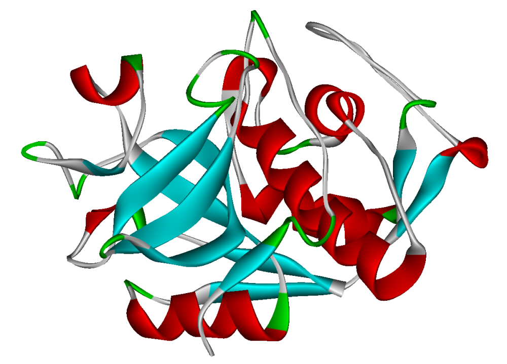
Cathepsins are proteases, which degrade proteins, found in all animals and other organisms. There are about a dozen members of this family distinguished by their structure, catalytic mechanism, and which proteins they cleave.
What are Cathepsins?
Cathepsins are proteases, which degrade proteins, found in all animals and other organisms. There are about a dozen members of this family distinguished by their structure, catalytic mechanism, and which proteins they cleave.
When do most members become activated? They are activated at low pH found in lysosomes.
Where does Cathepsin activity take place? Almost entirely within lysosome organelles, except for cathepsin K, which operates extracellularly after osteoclast secretion in bone resorption (type of cellular turnover).
Why are Cathepsins important? They play vital roles in mammalian cellular turnover and degrade polypeptides. They are distinguished by their substrate specificities.
What are the three types of Cathepsins?
The three types of proteases are listed in the table below:
What are the clinical significances of these Cathepsins?
Some of the most recent ones are listed in the table below.
| Cathepsin |
Clinical Significance |
A
 |
- Deficiencies in this protein are linked to multiple forms of galactosialidosis, a lysosomal storage disease ((Kleijer, 1996).
- Cathepsin A activity in lysates of metastatic lesions of malignant melanoma is significantly higher than in primary focus lysates (Kozlowski, 2000).
- Increased Cathepsin A in muscles moderately affected by muscular dystrophy and denervating diseases (Kar, 1976).
|
B
 |
- Cathepsin B in Alzheimer's studies have shown to attribute to beta-amyloid(Aβ) production by cleaving wild-typeβ-secretase site sequence in AβPP to produce Aβ. Cathepsin B inhibitors given to animal models with AβPP with the wild-type exhibited a reduction of Aβ in a manner consistent with β-secretase inhibition (Hook, 2011).
- Overexpression of the encoded protein, which is a member of the peptidase C1 family, has been associated with esophageal adenocarcinoma and other tumors (Homo Sapiens, 2016)
- Cathepsin B was also implicated in the progression of various human tumors[1] including ovarian cancer (Nomura, 2005).
- Cathepsin B is involved in apoptosis and degradation of myofibrillar proteins in myocardial infarction (Ge, 2006).
|
D |
- Cathepsin D (an aspartyl protease) appears to cleave a variety of substrates such as fibronectin and laminin. Unlike some of the other cathepsins, cathepsin D has some protease activity at neutral pH (Lkhide, 2004).
- High levels of this CtsD in tumor cells seems to be associated with greater invasiveness (Gong-jun, 2013)
|
|
K
 |
- Cathepsin K is the most potent mammalian collagenase and is involved in osteoporosis, a disease in which a decrease in bone density causes an increased risk for fracture. Osteoclasts are the bone resorbing cells of the body. They secrete cathepsin K in order to break down collagen, the major component of the non-mineral protein matrix of the bone (Shi, 1995)
- Cathepsin K, among others, plays a role in cancer metastasis through the degradation of the extracellular matrix (Gocheva, 2007).
- The genetic knockout for cathepsin S and K in mice with atherosclerosis was seen to reduce sizes of atherosclerotic lesions (Lutgens, 2006)
- Expression of cathepsin K in cultured endothelial cells is regulated by shear stress (Platt, 2007).
- Cathepsin K plays a role in arthritis (Salminen-Mankonen, 2007).
|
|
L
 |
- Mouse cathepsin L is homologous to human cathepsin V (Brömme, 1999) Its been shown to play a role in adipogenesis and glucose intolerance in mice. It degrades fibronectin, insulin receptor (IR), and insulin-like growth factor 1 receptor (IGF-1R). Cathepsin L-deficient mice were seen to have less adipose tissue, lower serum glucose and insulin levels, more insulin receptor subunits, more glucose transporter (GLUT4) and more fibronectin than wild type controls (Yang, 2007)
|
How were Cathepsins discovered?
A 1949 article from the Journal of Biological Chemistry has the earliest record of cathepsin research by Mary E. Maver and Antoinette E. Greco called The Hydrolysis of Nucleoproteins by Cathepsins from Calf Thymus. However, the references in the article show previous cathepsins were identified around the turn of the 20th century. Earlier work was completed by the laboratory of Max Bergmann, defining proteases in the first several decades of the century.
Related:
References
Brömme D, Li Z, Barnes M, Mehler E (February 1999). "Human cathepsin V functional expression, tissue distribution, electrostatic surface potential, enzymatic characterization, and chromosomal localization". Biochemistry 38 (8): 2377–85.doi:10.1021/bi982175f. PMID 10029531.
Kar, N. C., & Pearson, C. M. (1976). Arylamidase and Cathepsin-A Activity of Normal and Dystrophic Human Muscle. Experimental Biology and Medicine,151(3), 583-586. doi:10.3181/00379727-151-39264
Kleijer, W. J., Geilen, G. C., Janse, H. C., Diggelen, O. P., Zhou, X. Y., Galjart, N. J., . . . D'azzo, A. (1996). Cathepsin A Deficiency in Galactosialidosis: Studies of Patients and Carriers in 16 Families. Pediatr Res Pediatric Research, 39(6), 1067-1071. doi:10.1203/00006450-199606000-00022
Kozlowski, L., Wojtukiewicz, M. Z., & Ostrowska, H. (2000). Cathepsin A activity in primary and metastatic human melanocytic tumors. Archives of Dermatological Research, 292(2-3), 68-71. doi:10.1007/s004030050012
Ge, J., Zhao, G., Chen, R., Li, S., Wang, S., Zhang, X., . . . Yang, Y. (2006). Enhanced myocardial cathepsin B expression in patients with dilated cardiomyopathy.European Journal of Heart Failure, 8(3), 284-289. doi:10.1016/j.ejheart.2005.09.004
Gong-Jun Tan, Zheng-Ke Peng, Jin-Ping Lu, Fa-Qing Tang (2013). Cathepsins mediate tumor metastasis. World J Biol Chem. 2013 November 26; 4(4): 91–101. Published online 2013 November 26. doi: 10.4331/wjbc.v4.i4.91PMCID: PMC3856311
Homo sapiens complex locus CTSB, encoding cathepsin B. (n.d.). Retrieved May 18, 2016, from http://www.ncbi.nlm.nih.gov/IEB/Research/Acembly/av.cgi?db=human
Hook, G., Yu, J., Kindy, M. S., & Hook, V. (2014). Cysteine Protease Inhibitor, E64D, Of Cathepsin B Reduces Pglu-Abeta And Abeta, And Improves Memory Deficits In The Applon Mouse Model Of Ad. Alzheimer's & Dementia, 10(4). doi:10.1016/j.jalz.2014.05.638
Gocheva V, Joyce JA (January 2007). "Cysteine cathepsins and the cutting edge of cancer invasion". Cell Cycle 6 (1): 60–4.doi:10.4161/cc.6.1.3669. PMID 17245112.